Lung Cancer

Lung cancer is the most common cancer worldwide, with an estimated 1.8 million cases annually.

Although lung cancer is largely attributable to smoking cigarettes, or secondary exposure to tobacco smoke, the number of cases of lung cancer remains high in the developed world and is rising rapidly in the developing economies as affluence makes tobacco products more affordable. As a result, lung cancer is likely to remain a significant cause of death worldwide for the foreseeable future. Five-year survival for patients with lung cancer is 15%, meaning that there is ample opportunity for new treatments to improve the overall prognosis of lung cancer patients.

Approximately 208,000 people are diagnosed with lung cancer in the United States annually and approximately 158,000 people die from lung cancer each year.

Between 80 to 90% of lung cancer cases are caused by smoking tobacco. Lung cancer accounts for 14% of all cases of cancer in the US annually.1 Overall 5-year survival for patients with lung cancer is 15%.2 Diagnosis is generally based on chest X-ray and treatment, while generally determined by the type of lung cancer, usually involves both radiation therapy and chemotherapy.

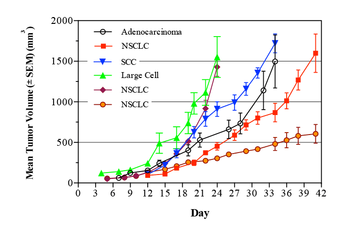
Lung cancer is broadly classified into two main types based on the cancer’s appearance under a microscope: non-small cell lung cancer (NSCLC) and small cell lung cancer (SCLC). Non-small cell lung cancer accounts for 80% of lung cancers, while small cell lung cancer accounts for the remaining 20%.3

NSCLC is further divided into four different types:

  • Squamous cell carcinoma. (SCC) also known as epidermoid carcinoma. The most common type of NSCLC squamous cell carcinoma forms in the lining of the bronchial tubes.
  • Adenocarcinoma. Adenocarcinoma is the most common type of lung cancer in women and in nonsmokers, adenocarcinoma forms in the mucus-producing glands of the lungs.
  • Bronchio-alveolar carcinoma. This type of lung cancer is a relatively rare adenocarcinoma that forms near the lungs’ air sacs.
  • Large-cell undifferentiated carcinoma.This rapidly growing cancer is composed of large cells forming an undifferentiated carcinoma typically near the outer edges or surfaces of the lungs.

Treatment for NSCLC typically involves surgery4 often with adjuvant chemotherapy usually platinum based (cisplatin, carboplatin), or taxanes (docetaxel, paclitaxel) and cytotoxic agents (gemcitabine, etoposide).5-7

Treatment for SCLC is generally by radiation and/or chemotherapy and does not commonly involve surgery.3 Chemotherapy usually consists of cisplatin and/or etoposide and combinations of these two compounds with paclitaxel, gemcitabine, vinorelbine and topotecan or irinotecan are also commonly used.8-10

Numerous alterations to oncogene expression have been reported in lung cancers.
As in many malignant tumors, lung cancers show a high degree of genetic instability. Activating mutations in the KRAS gene are seen in 25-40% of lung cancers . Approximately 3% of lung cancer patients have activating mutations in Braf, while 2% have activating mutations in the MEK/MAPK-1 gene, which lies in the same pathway. Both Braf and MEK lie in the Ras oncogene pathway, downstream of ras.

Reports indicate that 10-15% of lung cancers have activating mutations of the epidermal growth factor receptor (EGFR),11 while 2-4% of cases have activating mutations of the related gene HER2. Activations of the Ras and EGFR/Her-2 are almost invariably mutually exclusive, suggesting that each serve as driver mutations for the cancer, rather than incidental mutations that have occurred during the development of the tumor. Mutations in the Met and ALK oncogenes are found in relatively small percentages of patients (1%-7%) and are generally not found in in patients with activations of KRAS or EGFR/Her-2. Activation of the PI3K/AKT/mTOR pathway is seen in approximately 60% of NSCLC cases, but is generally seen in conjunction with either KRAS or EGFR/Her-2 activation and is thought not to be a primary driver of tumor growth. Additionally, mutations have been identified in the Ros-1, RET, FGFR1, and DDR2 oncogenes, as well as numerous tumor suppressor genes, but the exact incidence and role of these mutations in the development of lung cancer is still under investigation.

Animal Models of Lung Cancer

Pharma Models LLC offers several models of the different types of lung cancer. Most commonly used are the sub-cutaneous human tumor xenografts, and some of the cell lines used in these models can also be implanted orthotopically into the lungs of immune-compromised mice. Some cell lines can be implanted directly into common metastatic sites for human lung cancer, such as the brain and liver.

Subcutaneous Xenograft Models for Human Lung Cancer

There are a number of cell lines that are commonly used as subcutaneous xenograft models of human lung cancer in immunocompromised mice. Several growth curves are shown in Figure 1. In this example, four lung cancer cell lines were implanted subcutaneously in nude mice and the growth of the resulting tumors followed. In this model, treatment would typically begin at a mean tumor volume of 100 mm3, and continue until the negative control tumors reach a volume of 1500 mm3.

Figure 1
Figure 1: Representative Growth Curves for six Lung Cancer Cell Lines. Nude mice were implanted subcutaneously with approximately 2×10⁶ cells on Day 0. NSCLC = Non-small cell lung carcinoma. SCC= Squamous cell carcinoma.

Orthotopic Models of Lung Cancer

In addition to subcutaneous xenograft models of cancer, cell lines can also be implanted into the lungs of mice as an orthotopic model of lung cancer. Pharma Models LLC has luciferase expressing versions of many of the most commonly used human lung cancer cell lines that can be implanted by intrathoracic injection and monitored by quantifying luciferase expression. An example of the growth of an orthotopic lung cancer model is shown in Figure 2.

Figure 2

Figure 2

Figure 2: Orthotopic Lung Cancer in Nude Mice. Mice were implanted intrathoracically with 1×10⁶ luciferase expressing lung cancer cells. Mice were evaluated for luciferase expression in a Bruker in vivo Xtreme imaging system on Days 9, 16, 23 and 37 post tumor cell implantation. Image shows luminescence overlaid on X-ray.

Metastatic models of Lung Cancer

The brain is a frequent site for metastatic lung cancer. Clinically, brain metastases have special relevance because they are potentially more likely to cause death more quickly than the primary lung tumor. Lung cancer metastases in mice can be modeled by injecting tumor cells intra-cranially and then following the progress of the disease through in vivo imaging, survival curves, and ultimately, histologically. With Luciferase labeled lung cancer cells, the tumor can be followed as it progresses, and the extent of tumor growth quantified. An example of the imaging possible is shown in Figure 3. Please contact us for more details of these models.

Figure 3

Figure 3

Figure 3: Metastatic Lung Cancer in Brain. Mice were implanted intracranially with 2×10⁴ luciferase expressing lung cancer cells. Mice were evaluated for luciferase expression in a Bruker in vivo Xtreme imaging system on Day 7 post tumor cell implantation. Image shows luminescence overlaid on X-ray. Tumor growth was visualized with D-luciferin 6, 13, 20, 27 and 37 days post injection.

Syngeneic Models of Lung Cancer

Syngeneic models of cancer allow drugs to be tested in systems with mice that have intact immune systems. Although there are generally fewer cell lines available, the protocol is basically the same as for a human cell line – the cell lines can be implanted subcutaneously, directly into the thoracic cavity, or the model can be run as a metastatic model. Please contact us for details of orthotopic and metastatic syngeneic lung cancer models.

Figure 4: Representative growth curve for syngeneic lung cancer model LL2. Murine lung cancer cells were implanted subcutaneously in C57/B6 mice and tumors measured three times per week.

Figure 4

Bibliography

  1. United States Cancer Statistics; 1999-2008 cancer incidence and mortality data.
  2. Collins, LG; Haines C, Perkel R, Enck RE (2007). “Lung cancer: diagnosis and management”. American Family Physician (American Academy of Family Physicians) 75 (1): 56–63.
  3. Horn, L; Pao W, Johnson DH (2012). “89”. Harrison’s Principles of Internal Medicine (18th ed.). McGraw-Hill.
  4. Strand, TE; Rostad H, Damhuis RA, Norstein J (Jun 2007). “Risk factors for 30-day mortality after resection of lung cancer and prediction of their magnitude”. Thorax (BMJ Publishing Group Ltd.) 62 (11): 991–7. doi:10.1136/thx.2007.079145. PMC 2117132. PMID 17573442.
  5. Mehra R, Treat J (2008). Fishman’s Pulmonary Diseases and Disorders (4th ed.). McGraw-Hill. p. 1876.
  6. Clegg, A; Scott DA, Hewitson P et al. (January 2002). “Clinical and cost effectiveness of paclitaxel, docetaxel, gemcitabine, and vinorelbine in non-small cell lung cancer: a systematic review”. Thorax 57 (1): 20–28.
  7. Fuld AD, Dragnev KH, Rigas JR (J2010). “Pemetrexed in advanced non-small-cell lung cancer”. Expert Opin Pharmacother 11 (8): 1387–402.
  8. Murray, N; Turrisi AT (2006). “A review of first-line treatment for small-cell lung cancer”. Journal of Thoracic Oncology 1 (3): 270–278.
  9. Azim, HA; Ganti AK (2007). “Treatment options for relapsed small-cell lung cancer”. Anticancer drugs 18 (3): 255–261.
  10. MacCallum, C; Gillenwater HH (2006). “Second-line treatment of small-cell lung cancer”. Current Oncology Reports 8 (4): 258–264.
  11. Giard, DJ; Aaronson, SA; Todaro, GJ; Arnstein, P; Kersey, JH; Dosik, H; Parks, WP (1973). “In vitro cultivation of human tumors: Establishment of cell lines derived from a series of solid tumors”. Journal of the National Cancer Institute 51 (5): 1417–23.
  12. Ledinko N, Costantino RL. Modulation of p53 gene expression and cytokeratin 18 in retinoid-mediated invasion suppressed lung carcinoma cells. Anticancer Res. 1990;10:1335-9.
Contact Us
Cancer
Inflammatory Diseases